About Medical Pathological Microscopes
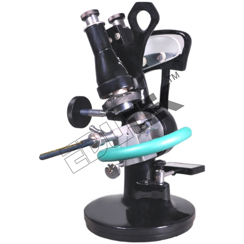
Medical Pathological Microscopes ETMS-132AMedical Pathological Microscopes (Advanced)SPECIFICATIONS- Viewing Head: Monocular 45
- Eyepiece : WF 10x-18mm
- Nosepiece: Quadruple, Upright
- Objective: Achro 4x,10x, 40x(SL) and 100x(SL)
- Mechanical Stage: 120x120mm with XY movement
- Focusing Movement: Separate Coarse and Fine movement
- Condenser: Moveable Abbe N A 1.25 with Iris Diaphragm
- Light Source: Plano-concave mirror, LED Lamp
- Filter: Blue
- Carrying Case: Styrofoam Packing
- Supplied with : Manual & Dust cover
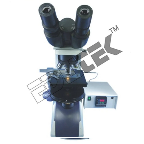
Magnification for Detailed AnalysisWith a magnification range from 40X to 1500X, this microscope covers a diverse spectrum of pathological and medical investigations. Achromatic objectives and wide-field eyepieces provide clear, distortion-free images, making it suitable for examining cells, tissues, and microorganisms with high fidelity. Precise focusing is achieved through coaxial coarse and fine adjustments.
User-Friendly and Versatile OperationEngineered for ease of use, the microscope features a 360 rotatable head and an inclined eyepiece tube, accommodating comfortable viewing angles. The double-layer mechanical stage offers precise X-Y movement over 75 x 40 mm, ensuring smooth sample navigation. Diopter and interpupillary adjustments enable personalized settings for each user.
Robust Construction with Enhanced OpticsCrafted from rigid, corrosion-resistant metal with anti-fungal coated optics, this instrument promises reliability and long life even in demanding environments. A swing-out filter holder, adjustable LED or halogen illumination, and a dust cover help maintain performance and cleanliness. Spare parts like lamps and eyepieces are readily available, supporting ongoing use.
Seamless Digital Imaging and DocumentationThe integrated trinocular drawtube allows for straightforward attachment of digital cameras, offering high-sensitivity CMOS imaging at up to 1920 x 1080 resolution. Images and videos can be captured in JPEG or PNG formats, facilitating easy documentation and remote sharing in lab or clinical settings. Full HD video and up to 5 MP still image capture support detailed analysis.
FAQs of Medical Pathological Microscopes:
Q: How does the quadruple ball bearing nosepiece benefit users during specimen examination?
A: The quadruple ball bearing nosepiece allows smooth and reliable rotation between four objective lenses, enabling swift changes in magnification. This ensures seamless workflow and precise adjustments without disturbing the focus or alignment, enhancing productivity during examination.
Q: What types of samples and applications is this microscope suitable for?
A: This microscope is ideal for medical pathology, biological research, and educational use. With its 40X1500X magnification range and precision mechanics, it supports examination of tissue sections, blood smears, bacteria, and other cellular or microbial specimens.
Q: When should the diopter adjustment be used, and what is its purpose?
A: The diopter adjustment on the left eyepiece allows users to compensate for differences in vision between their eyes. It should be adjusted when first using the microscope to obtain a sharp image in both eyes, improving comfort and accuracy during extended use.
Q: Where can digital imaging be applied with this microscope?
A: Digital imaging is facilitated via the trinocular port and USB 2.0 interface, enabling integration with computers and display systems. This feature is especially valuable in laboratories, hospitals, educational institutions, or telemedicine settings for documentation and collaborative analysis.
Q: What is the process for attaching a camera, and what image quality can be expected?
A: Attaching a camera is straightforward via the trinocular drawtube. Once connected, high-sensitivity CMOS sensors capture images at up to 1920 x 1080 pixels for video (30fps) and stills up to 5 MP, depending on the camera used. This ensures detailed, high-resolution documentation of specimens.
Q: How does the microscopes illumination system improve specimen visibility?
A: The microscope offers both LED and halogen illumination with adjustable intensity, ensuring optimal lighting for transmitted and reflected observation. This flexibility allows users to visualize specimens with enhanced clarity and contrast under various conditions.
Q: What are the main benefits of the microscopes construction and optical features?
A: Key benefits include a sturdy, corrosion-resistant metal frame, anti-fungal coated optics, dust protection, and a mechanical double-layer stage. These features ensure reliable operation, long-term resistance to environmental factors, and clear, distortion-free imaging for accurate analyses.


 Send Inquiry
 Send Inquiry
