


Malaria Detection Microscopes
Product Details:
X
Product Description
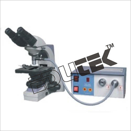
Malaria Detection Microscopes ETMS-144This a new method for identifying the malaria parasite in the peripheral blood. It involves staining of the centrifuged and compressed red cell layer with acridine orange and its examination under UV light source. It is fast, easy and claimed to be more sensitive than the traditional thick smear examination.
Method: The tube is a high-precision glass hematocrit tube, pre-coated internally with acridine orange stain and potassium oxalate. It is filled with 55-65 micro liters of blood from a finger, ear or heel puncture. A clear plastic closure is then attached. A precisely made cylindrical float, designed to be suspended in the packed red blood cells, is inserted. The tube is centrifuged at 12,000 rpm for 5 minutes. The components of the buffy coat separate according to their densities, forming discrete bands. Because the float occupies 90% of the internal lumen of the tube, the leukocyte and the thrombocyte cell band widths and the top-most area of red cells are enlarged to 10 times normal. The tube is placed on the tube holder and examined using a standard white light microscope equipped with the UV microscope adapter, an epi-illuminated microscope objective. Fluorescing parasites are then observed at the red blood cell/white blood cell interface. The key feature of the method is centrifugation and thereby concentration of the red blood cells in a predictable area of the tube, making detection easy and fast. Red cells containing Plasmodia are less dense than normal ones and concentrate just below the leukocytes, at the top of the erythrocyte column. The float forces all the surrounding red cells into the 40 micron space between its outside circumference and the inside of the tube. Since the parasites contain DNA which takes up the acridine orange stain, they appear as bright specks of light among the non-fluorescing red cells. Virtually all of the parasites found in the 60 microliter of blood can be visualized by rotating the tube under the microscope. A negative test can be reported within one minute and positive result within minutes. is a new method for identifying the malarial parasite in the peripheral blood. It involves staining of the centrifuged and compressed red cell layer with acridine orange and its examination under UV light source. It is fast, easy and claimed to be more sensitive than the traditional thick smear examination.
SPECIFICATIONS
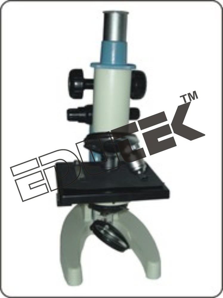
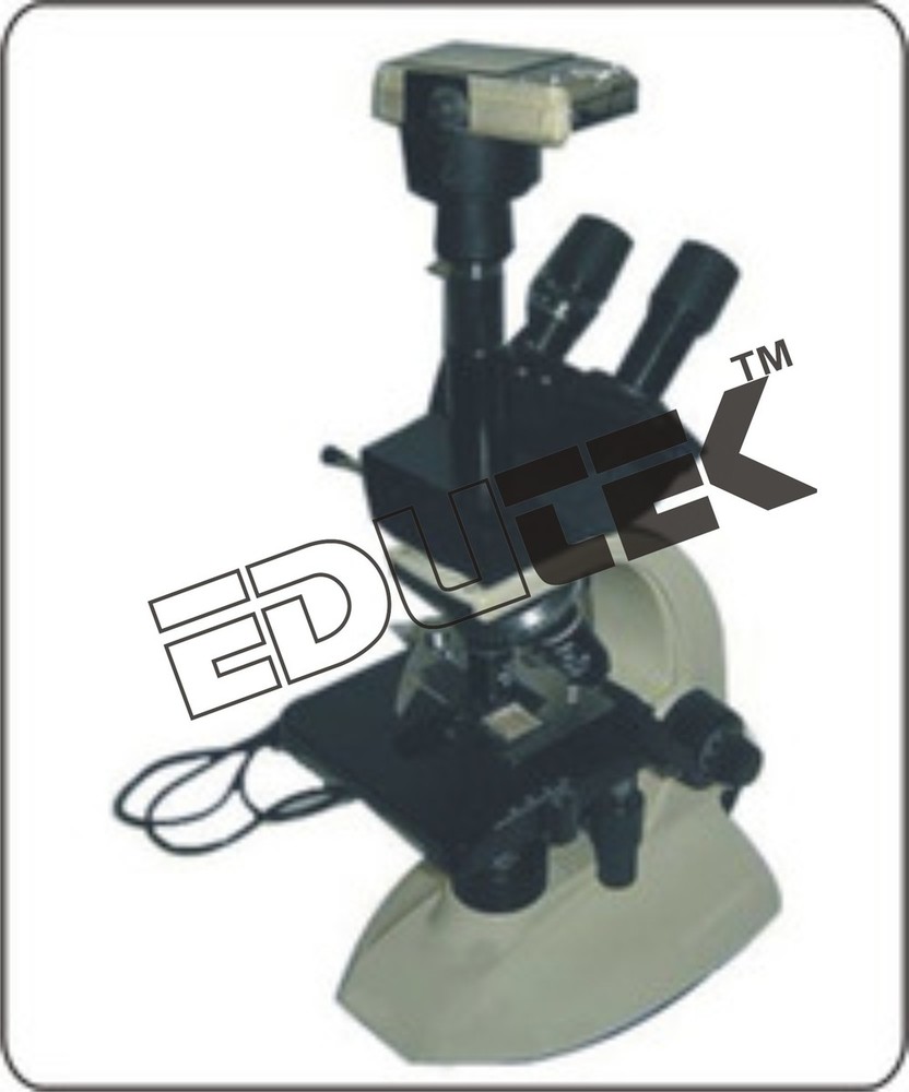
- Coaxial Binocular Microscope,
- UV adapter
- Excitation Filter D470/40x -- 8 mm dia, 3 mm thick (Excitation (x) filter should be positioned with the arrow pointing toward the specimen, toward the inside of the cube, and away from the light source.),
- Dichroic Beam Splitter 500 DCLP, 7.8x10.95x0.75mm (Dichroic mirrors should be mounted with the coated surface towards the light source, excitation filters, and the specimen).
- Barrier Filter E5151PV2, 9.90mm dia, 3mmthick (Emission (m) filters should be placed with the arrow pointing towards the specimen, towards the inside of the cube, and away from the detector / eye),
- 62x objective (gives sharp image of plasmodium vivax, phalciparum and shizhont etc),
- Focusing lens Passes white light from illuminator to the excitation filter.(size 13mm or above),
- Standard 36 TPI Threading Assures adaptability to all.
Enter Buying Requirement Details
Other Products in 'Research Microscope' category

"We deal all over World but our main domestic market is South India"

- EDUTEK INSTRUMENTATION
None - Factory: 70, Edutek House, Main Road Vikaspuri, Industrial Area, Ambala Cantt - 133006, Haryana, India
- Phone :91-171-2699855

- Email Id : edutekgroup@gmail.com
- Mr Aditya Kumar (Director-International Sales)
- Mobile :+919996644855, +918930444855
- edutekgroup@gmail.com
 |
EDUTEK INSTRUMENTATION
All Rights Reserved.(Terms of Use) Developed and Managed by Infocom Network Private Limited. |