EDUTEK INSTRUMENTATION
GST : 06AEWPJ6896D1ZU

call images

Call us

07313726362

Fluorescent Microscope

Fluorescent Microscope

Price 1000.0 INR/ Piece

MOQ : 1 Piece

Fluorescent Microscope Specification

  • Spare Parts
  • Replacement filters, additional bulbs, objective lenses
  • Material
  • Mild Steel
  • View Head
  • Trinocular 30 inclined, 360 rotatable
  • Features
  • Sharp fluorescence imaging, ergonomic design, anti-fungal optics, built-in camera for documentation
  • Application
  • Research Microscope
  • Usage
  • Laboratory
  • Product Type
  • Fluorescent Microscope
  • Focus System
  • Coaxial coarse and fine focusing mechanism
  • Theory
  • Fluorescence Microscope
  • Drawtube
  • Other
  • Sensor
  • CMOS sensor, High sensitivity for fluorescence
  • Resolution
  • 1920 x 1080 pixels (Full HD)
  • Interface
  • USB 2.0, HDMI
  • Frame Rate
  • 30fps (real-time observation)
  • Focal Distance
  • 50mm - 75mm (adjustable) Millimeter (mm)
  • Magnification
  • 40x to 1000x (with objective lenses)
  • Dimensions
  • Approx. 400 x 300 x 480 mm Millimeter (mm)
  • Focus Range
  • 30mm (coarse), 0.002mm (fine) Millimeter (mm)
  • Eyepieces
  • Widefield WF10x/20mm or WF15x optional
  • Eyepiece Tube
  • Interpupillary distance adjustable, 48-75mm
  • Illumination
  • LED Fluorescence Illumination with filter cube system
  • Coarse Adjustment Range
  • 30mm
  • Fine Adjustment Range
  • 0.002mm precision
  • Working Stage
  • Double layer mechanical stage, size 140 x 140mm, travel 75 x 50 mm Square Millimeter (mm2)
  • Still Image Capture Resolution
  • Up to 5MP digital capture
  • Video Capture Resolution
  • Full HD 1080p video recording
  • Image Format
  • JPEG, BMP, TIFF
  • Interpupillary Distance
  • 48-75mm adjustable Millimeter (mm)
  • Objective Achromatic
  • 4x, 10x, 40x(S), 100x(S, Oil) Anti-fungal, strain-free Achromatic objectives
  • Condenser
  • Abbe condenser N.A. 1.25 with iris diaphragm & filter holder
  • Light Source
  • High-intensity LED (blue, green filters for fluorescence), 3W, long life
  • Relative Humidity
  • Maximum 85% non-condensing
  • Dust Cover
  • Included for instrument protection
  • Operating Temperature
  • 5°C to 40°C
  • Packaging
  • Foam-protected in sturdy carton for safe transport
  • Body Construction
  • Sturdy metal base with anti-vibration pads
  • Adjustment Knobs
  • Ergonomically placed coaxial knobs for easy operation
  • Stage Movement
  • Low-position coaxial controls for X/Y movement
  • Power Supply
  • Universal 100-240V AC, 50/60Hz
  • Filter Sets
  • Blue, Green, UV filter cubes included
 

Fluorescent Microscope Trade Information

  • Minimum Order Quantity
  • 1 Piece
  • Payment Terms
  • Cheque, Cash in Advance (CID), Telegraphic Transfer (T/T), Letter of Credit (L/C), Cash Advance (CA)
  • Sample Available
  • Yes
  • Sample Policy
  • Sample costs shipping and taxes has to be paid by the buyer
  • Packaging Details
  • carton box, export quality also available
  • Main Export Market(s)
  • Asia, Eastern Europe, Western Europe, Middle East, Africa
  • Main Domestic Market
  • All India
  • Certifications
  • ISO 9001, 14001 ,CE
 

About Fluorescent Microscope
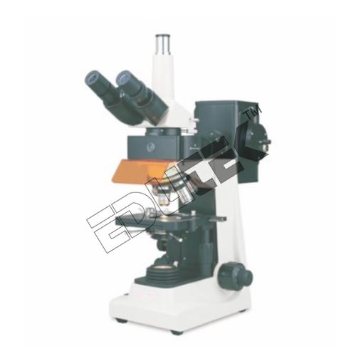
Fluorescent Microscope ETMS-143

Fluorescent Microscope is used for capillary examination. This technique is a new, highly sensitive rapid method of diagnosing malaria. It is also useful in the diagnosis of filariasis and leptospirosis. The principle of this method is the malaria parasite picks up fluorescent stain into their nucleus and cytoplasm, so that its morphologic characteristics can be examined by fluorescent microscopy. The nucleus appears green and the cytoplasm reddish orange,

 The Capillary is coated with Acridine orange on its inside. Parents Blood is loaded into this capillary and centrifuged at 12000 rpm for5 minutes. The Blood components settle at different levels in the capillary. These different layers are displayed in the picture.

 Examination of the area just below granulocyte-RBC junction helps to detect the malarial parasites, as these are concentrated in this are upto 1000 times. Examination of Lymphocyte / monocyte layer using transmitted light helps to detect the schizonts, and gametocytes which gets concentrated in this layer very easily.

 Method of Examination:

 The granulocyte layer is then seen under 62x oil objective. By changing the light source to fluorescent light the parasitic forms can be seen as greenish orange stained structures. Using only transmitted light the parasitic forms are seen as block pigmented structures as shown in the preceding photomicrographs.
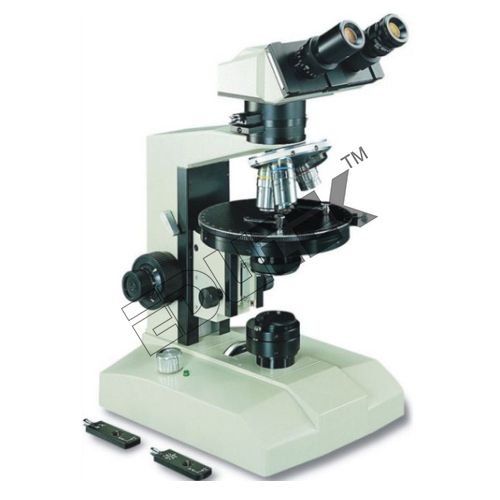

Exceptional Optical Clarity

Equipped with anti-fungal, strain-free achromatic objectives from 4x to 100x (oil), this microscope delivers sharp and clear fluorescence images. The Abbe condenser with iris diaphragm and filter holder ensures optimal light focusing, allowing for detailed observation of cellular structures in research and diagnostics.


Ergonomic and User-Friendly Operation

With coaxial coarse and fine focusing, low-position X/Y movement controls, and wide interpupillary adjustment (4875 mm), this microscope prioritizes usability. The trinocular head is inclined at 30 for comfortable prolonged viewing and rotates 360 to fit varied laboratory settings and preferences.
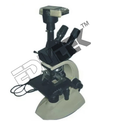

Advanced Imaging and Documentation

The integrated high-sensitivity CMOS sensor allows for Full HD live viewing and documentation. Users can capture JPEG, BMP, or TIFF images (up to 5MP) and record 1080p video directly, making the system ideal for archiving results, sharing findings, or remote demonstrations.

FAQs of Fluorescent Microscope:


Q: How does the microscopes fluorescence feature benefit cellular studies?

A: Fluorescence microscopy exploits the property of certain specimens to emit light when excited by specific wavelengths. This allows the observation and detailed study of cellular structures, proteins, and components with exceptional contrast and specificity compared to traditional brightfield microscopy.

Q: What is the process for capturing images and videos using the built-in camera?

A: The built-in CMOS camera supports Full HD resolution and connects via USB 2.0 or HDMI. Simply adjust the focus, select your preferred view, and use included software to capture still images up to 5MP or record 1080p video at 30fps, making it easy to document and share results.

Q: When should I use different filter cubes (blue, green, UV) during fluorescence imaging?

A: Select filter cubes based on the fluorescent dyes or markers applied to your samples. Blue, green, and UV filters are included to match common fluorophores, ensuring optimal excitation and emission for clear visualization of distinct cellular targets.

Q: Where is this fluorescent microscope ideally used?

A: This system is suitable for educational institutions, clinical laboratories, and research facilities that require precise fluorescence imaging and documentation capabilities. Its robust construction and ergonomic design make it versatile for regular laboratory use in varied environmental conditions.

Q: What are the ergonomic features that enhance ease of use?

A: The microscope includes low-position coaxial stage controls, ergonomically-placed focus knobs, and a 30 inclined, 360 rotatable trinocular head. The interpupillary distance is widely adjustable (4875mm), ensuring comfort during extended observation sessions.

Q: How is the instrument protected during transportation and storage?

A: A foam-protected, sturdy carton ensures safe transport, while the included dust cover protects the microscope from contamination when not in use, preserving optical quality and longevity.

Q: What spare parts and accessories are available for continued usage?

A: Replacement filter cubes, additional bulbs, objective lenses, and other spare parts are available to ensure uninterrupted use and maintain instrument performance over time.

Tell us about your requirement
product

Price:

Quantity
Select Unit

  • 50
  • 100
  • 200
  • 250
  • 500
  • 1000+
Additional detail
Mobile number

Email

More Products in Research Microscope Category

Binocular Microscope

Binocular Microscope

Price 1000.0 INR / Piece

Minimum Order Quantity : 1 Piece

Coarse Adjustment Range : 15 mm travel approximately.

Focus System : Coarse and fine coaxial focusing system.

Interface : Not applicable (standard optical use); optional camera port available in some models.

Focus Range : Coarse and fine adjustments available; range typically 15 mm for coarse. Millimeter (mm)

Optional Accessories for Microscopes

Optional Accessories for Microscopes

Price 1000.0 INR / Piece

Minimum Order Quantity : 1 Piece

Coarse Adjustment Range : Up to 30mm depending on stand model

Focus System : Manual and motorized focus accessories available

Interface : Compatible with USB and HDMI interfaces

Focus Range : Enhanced with coarse and fine focus adjustment accessories

Medical Pathological Microscopes

Medical Pathological Microscopes

Price 1000.0 INR / Piece

Minimum Order Quantity : 1 Piece

Coarse Adjustment Range : 30 mm

Focus System : Coaxial coarse and fine focusing mechanism

Interface : USB 2.0 (when camera attached)

Focus Range : Up to 15mm Coarse/0.002mm Fine

Research Polarimeter

Research Polarimeter

Price 1000.0 INR / Piece

Minimum Order Quantity : 1 Piece

Coarse Adjustment Range : 30 mm

Focus System : Manual coaxial coarse and fine adjustment knobs

Interface : Manual readings, digital output (optional)

Focus Range : Coarse and fine focus


"We deal all over World but our main domestic market is South India"
Back to top